Diploschistes diacapsis (Ach.) Lumbsch
Lichenologist, 20: 20, 1988. Basionym: Urceolaria diacapsis Ach. - Syn. Meth. Lich.: 339, 1814.
Synonyms: Diploschistes albescens Lettau; Diploschistes albissimus (Ach.) Dalla Torre & Sarnth.; Diploschistes gypsaceus auct. p.p.; Diploschistes gypsaceus subsp. neutrophilus Clauzade & Cl. Roux; Diploschistes induratus (Vain.) Zahlbr.; Diploschistes minor (Kremp.) Zahlbr.; Diploschistes neutrophilus (Clauzade & Cl. Roux) Fern.-Brime & Llimona; Diploschistes ocellatus var. fallax Werner; Diploschistes scruposus subsp. albescens (Lettau) Clauzade & Cl. Roux; Diploschistes steppicus Reichert; Urceolaria scruposa var. diacapsis (Ach.) Nyl.; Urceolaria sicula Jatta?
Distribution: N - TAA (Nascimbene & al. 2022), Lomb, Piem (Isocrono & Falletti 1999, Morisi 2005), Emil (Nimis & al. 1996, Fariselli & al. 2020), Lig. C - Tosc (Pišút 1997). S - Camp, Bas (Nimis & Tretiach 1999), Cal (Puntillo & Puntillo 2004), Si (Pišút 1995).
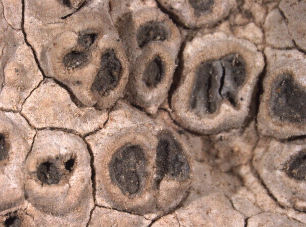
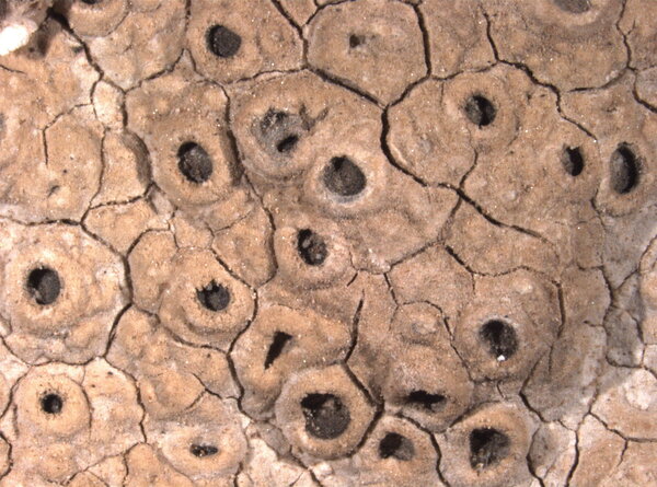
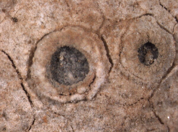
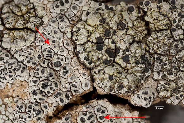
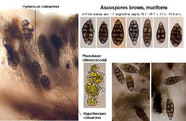
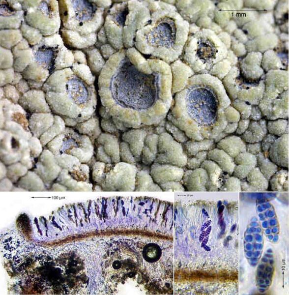
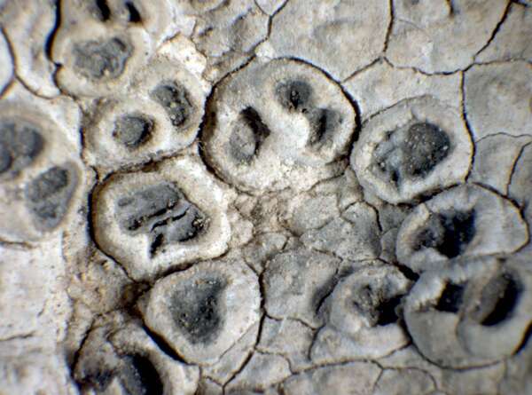
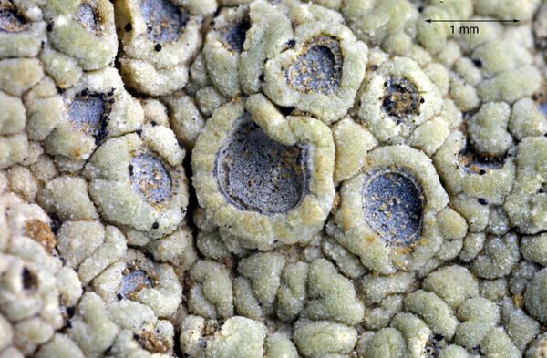
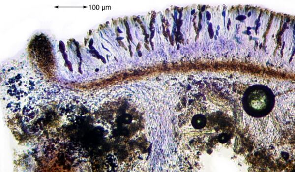
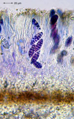
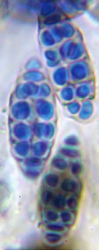
Description: Thallus crustose, episubstratic, rimose- to verrucose-areolate, whitish to grey, consisting of irregularly angular, 0.5-2.5 mm wide and 1-3 mm thick, flat to convex, dull, usually pruinose areoles. Medulla white, I- or rarely I+ blue. Apothecia lecanorine, urceolate, semi-immersed to sessile, up to 2.5 mm across, with a black, but often slightly grey-pruinose, concave disc, and a thick thalline margin. Proper exciple blackish brown, 60-80 µm wide laterally, pseudoparenchymatous; epithecium poorly differentiated, colourless to brownish; hymenium colourless, 110-180 µm high, non amyloid; paraphyses simple, flexuose, 1-2 µm thick, the apical cells not swollen; hypothecium colourless, c. 15 m high. Asci 4-8-spored, narrowly clavate to subcylindrical, the wall evenly thickened when mature, the somewhat abrupt apical thickening with a thin, internal apical beak, lacking any apical apparatus, the contents I+ orange-red, the walls I-, not fissitunicate. Ascospores muriform, with 3-6 transverse and 1-2 longitudinal septa, at first hyaline then turning brown, broadly ellipsoid, 20-40 x 9-17 µm; Pycnidia black, immersed in thallus, hyaline to brownish, cerebriform. Conidia bacilliform, 4-6 x 1-1.5 µm. Photobiont chlorococcoid. Spot tests: K- or K+ yellow to red, C+ red, KC-, P-, UV-. Chemistry: diploschistesic and lecanoric acids (both major) and orsellinic acid (minor).Note: a widespread species of arid grasslands, found on calciferous or base-rich soil, especially on gypsum, in open, dry situations; perhaps more widespread throughout the country. D. neutrophilus was originally segregated from D. diacapsis on account of its different ecology (it grows on neutral sandy to clay soil) and the amyloid reaction of the medulla, a character not confirmed by Fernández-Brime & al. (2013), although their molecular data indicated that this, in spite of the very weak morphological differences, could be a distinct species, differing from the calcicolous D. diacapsis. However, data by Zhao & al. (2017) suggest that D. neutrophilus should best considered as a synonym of D. diacapsis.
Growth form: Crustose
Substrata: soil, terricolous mosses, and plant debris
Photobiont: green algae other than Trentepohlia
Reproductive strategy: mainly sexual
Subcontinental: restricted to areas with a dry-subcontinental climate (e.g. dry Alpine valleys, parts of Mediterranean Italy)
Commonnes-rarity: (info)
Alpine belt: absent
Subalpine belt: absent
Oromediterranean belt: absent
Montane belt: extremely rare
Submediterranean belt: very rare
Padanian area: absent
Humid submediterranean belt: rare
Humid mediterranean belt: rare
Dry mediterranean belt: rather rare
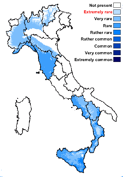
Predictive model
Herbarium samples

Felix Schumm – CC BY-SA 4.0
Image from: F. Schumm (2008) - Flechten Madeiras, der Kanaren und Azoren. Beck, OHG - ISBN: 978-3-00-023700-3

P.L. Nimis; Owner: Department of Life Sciences, University of Trieste
Herbarium: TSB (15119)
2001/11/24
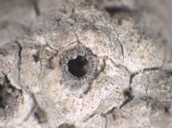

P.L. Nimis; Owner: Department of Life Sciences, University of Trieste
Herbarium: TSB (35712)
2003/01/22
young apothecium
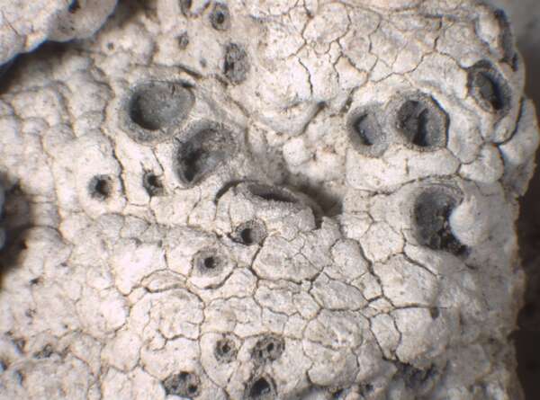

P.L. Nimis; Owner: Department of Life Sciences, University of Trieste
Herbarium: TSB (35712)
2003/01/22
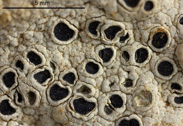

Felix Schumm – CC BY-SA 4.0
[16327], Mallorca (Süd): Zwischen San Marina und La Lova des Llatres, lichtoffener Straßenrand; 39,39117° N, 2,76329° E, 100 m. Leg. Schumm, Frahm & Lüth, 12.03.2010 (2), det. Schumm2010
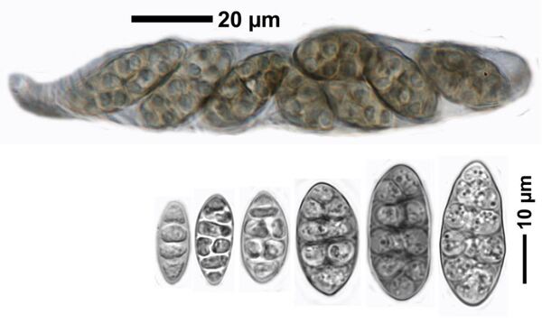

Felix Schumm – CC BY-SA 4.0
[16327], Mallorca (Süd): Zwischen San Marina und La Lova des Llatres, lichtoffener Straßenrand; 39,39117° N, 2,76329° E, 100 m. Leg. Schumm, Frahm & Lüth, 12.03.2010 (2), det. Schumm2010

Felix Schumm - CC BY-SA 4.0
Image from: F. Schumm (2008) - Flechten Madeiras, der Kanaren und Azoren. Beck, OHG - ISBN: 978-3-00-023700-3

Felix Schumm - CC BY-SA 4.0
Image from: F. Schumm (2008) - Flechten Madeiras, der Kanaren und Azoren. Beck, OHG - ISBN: 978-3-00-023700-3

Felix Schumm - CC BY-SA 4.0
Image from: F. Schumm (2008) - Flechten Madeiras, der Kanaren und Azoren. Beck, OHG - ISBN: 978-3-00-023700-3

Felix Schumm - CC BY-SA 4.0
Image from: F. Schumm (2008) - Flechten Madeiras, der Kanaren und Azoren. Beck, OHG - ISBN: 978-3-00-023700-3
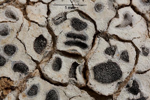
Ulrich Kirschbaum CC BY-SA 4.0 - Source: https://www.thm.de/lse/ulrich-kirschbaum/flechtenbilder
Canary Islands; La Gomera-M; e of Chipude: Top of the Fortaleza. On soil
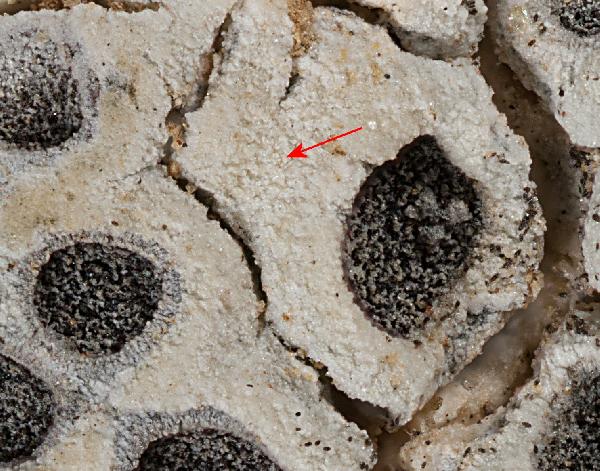
Ulrich Kirschbaum CC BY-SA 4.0 - Source: https://www.thm.de/lse/ulrich-kirschbaum/flechtenbilder
Canary Islands; La Gomera-M; e of Chipude: Top of the Fortaleza. On soil
Growth form: Crustose
Substrata: soil, terricolous mosses, and plant debris
Photobiont: green algae other than Trentepohlia
Reproductive strategy: mainly sexual
Subcontinental: restricted to areas with a dry-subcontinental climate (e.g. dry Alpine valleys, parts of Mediterranean Italy)
Commonnes-rarity: (info)
Alpine belt: absent
Subalpine belt: absent
Oromediterranean belt: absent
Montane belt: extremely rare
Submediterranean belt: very rare
Padanian area: absent
Humid submediterranean belt: rare
Humid mediterranean belt: rare
Dry mediterranean belt: rather rare

Predictive model
| Herbarium samples |


Felix Schumm – CC BY-SA 4.0
Image from: F. Schumm (2008) - Flechten Madeiras, der Kanaren und Azoren. Beck, OHG - ISBN: 978-3-00-023700-3


P.L. Nimis; Owner: Department of Life Sciences, University of Trieste
Herbarium: TSB (15119)
2001/11/24


P.L. Nimis; Owner: Department of Life Sciences, University of Trieste
Herbarium: TSB (35712)
2003/01/22
young apothecium


P.L. Nimis; Owner: Department of Life Sciences, University of Trieste
Herbarium: TSB (35712)
2003/01/22


Felix Schumm – CC BY-SA 4.0
[16327], Mallorca (Süd): Zwischen San Marina und La Lova des Llatres, lichtoffener Straßenrand; 39,39117° N, 2,76329° E, 100 m. Leg. Schumm, Frahm & Lüth, 12.03.2010 (2), det. Schumm2010


Felix Schumm – CC BY-SA 4.0
[16327], Mallorca (Süd): Zwischen San Marina und La Lova des Llatres, lichtoffener Straßenrand; 39,39117° N, 2,76329° E, 100 m. Leg. Schumm, Frahm & Lüth, 12.03.2010 (2), det. Schumm2010


Felix Schumm - CC BY-SA 4.0
Image from: F. Schumm (2008) - Flechten Madeiras, der Kanaren und Azoren. Beck, OHG - ISBN: 978-3-00-023700-3


Felix Schumm - CC BY-SA 4.0
Image from: F. Schumm (2008) - Flechten Madeiras, der Kanaren und Azoren. Beck, OHG - ISBN: 978-3-00-023700-3


Felix Schumm - CC BY-SA 4.0
Image from: F. Schumm (2008) - Flechten Madeiras, der Kanaren und Azoren. Beck, OHG - ISBN: 978-3-00-023700-3


Felix Schumm - CC BY-SA 4.0
Image from: F. Schumm (2008) - Flechten Madeiras, der Kanaren und Azoren. Beck, OHG - ISBN: 978-3-00-023700-3

Ulrich Kirschbaum CC BY-SA 4.0 - Source: https://www.thm.de/lse/ulrich-kirschbaum/flechtenbilder
Canary Islands; La Gomera-M; e of Chipude: Top of the Fortaleza. On soil

 Index Fungorum
 GBIF
GBIF
