Anema nummularium (Dufour ex Durieu & Mont.) Nyl. ex Forssell var. nummularium
N. Acta Reg. Soc. Sci. Upsal., ser. 3, 13, 6: 93, 1885. Basionym: Collema nummularium Dufour ex Durieu & Mont. in Durieu - Expl. Sci. Algérie., 1: 200, 1846.
Synonyms: Anema nummulariellum Nyl.; Omphalaria frustillata Nyl.; Tyrea nummularia (Durieu & Mont.) Zahlbr.
Distribution: N - VG, Frl, Ven (Lazzarin 2000b), TAA (Nascimbene & al. 2022), Lomb (Lazzarin 2000b, Brackel 2013), Piem (Matteucci & al. 2013), VA (Gazzano & al. 2009, 2009b), Emil (Fariselli & al. 2020), Lig (Lazzarin 2000b, Watson 2014). C - Tosc, Marc (Nimis & Tretiach 1999), Abr (Nimis & Tretiach 1999), Mol (Nimis & Tretiach 1999, Caporale & al. 2008), Sar. S - Camp (Aprile & al. 2003b, Nimis & Tretiach 2004, Garofalo & al. 2010), Pugl, Bas (Bartoli & Puntillo 1998), Cal (Puntillo 1996), Si (Nimis & al. 1994, Ottonello & al. 1994, Ravera & al. 2023b).
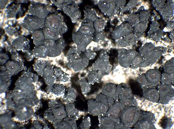
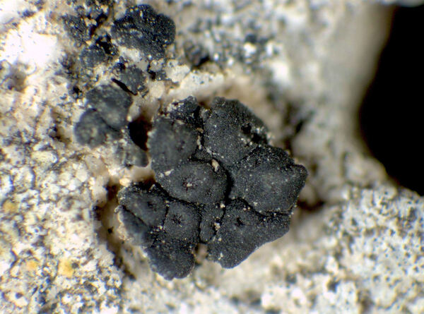
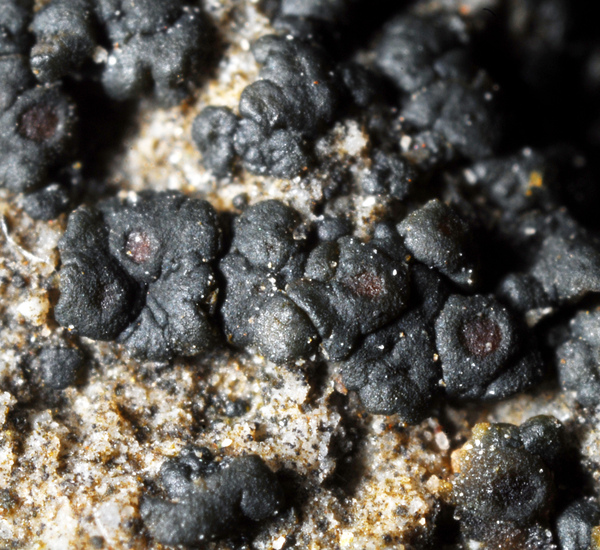
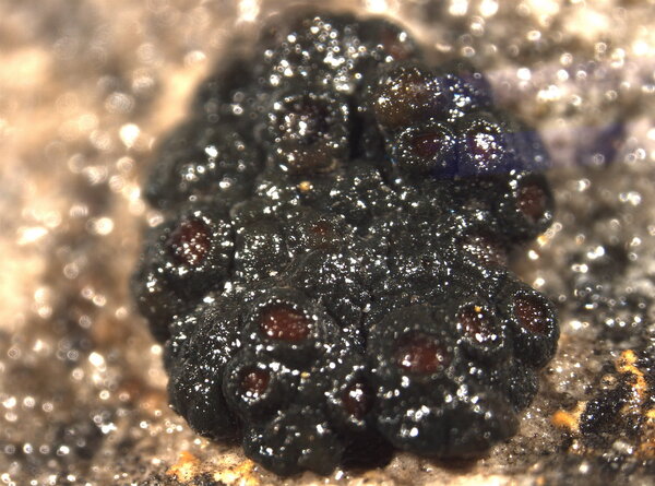
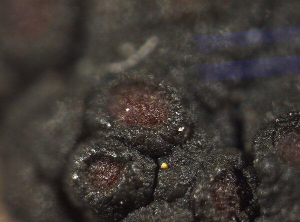
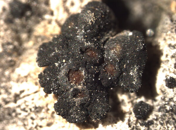
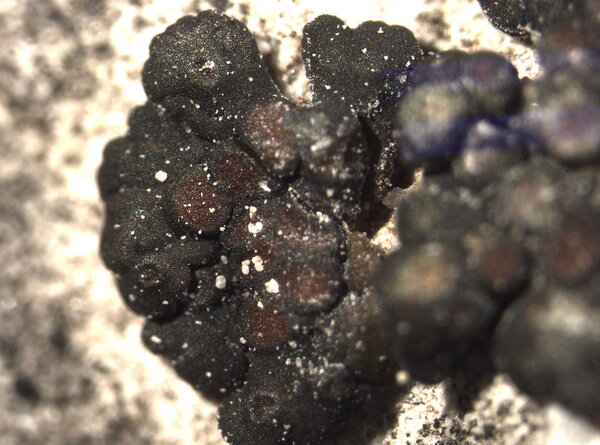
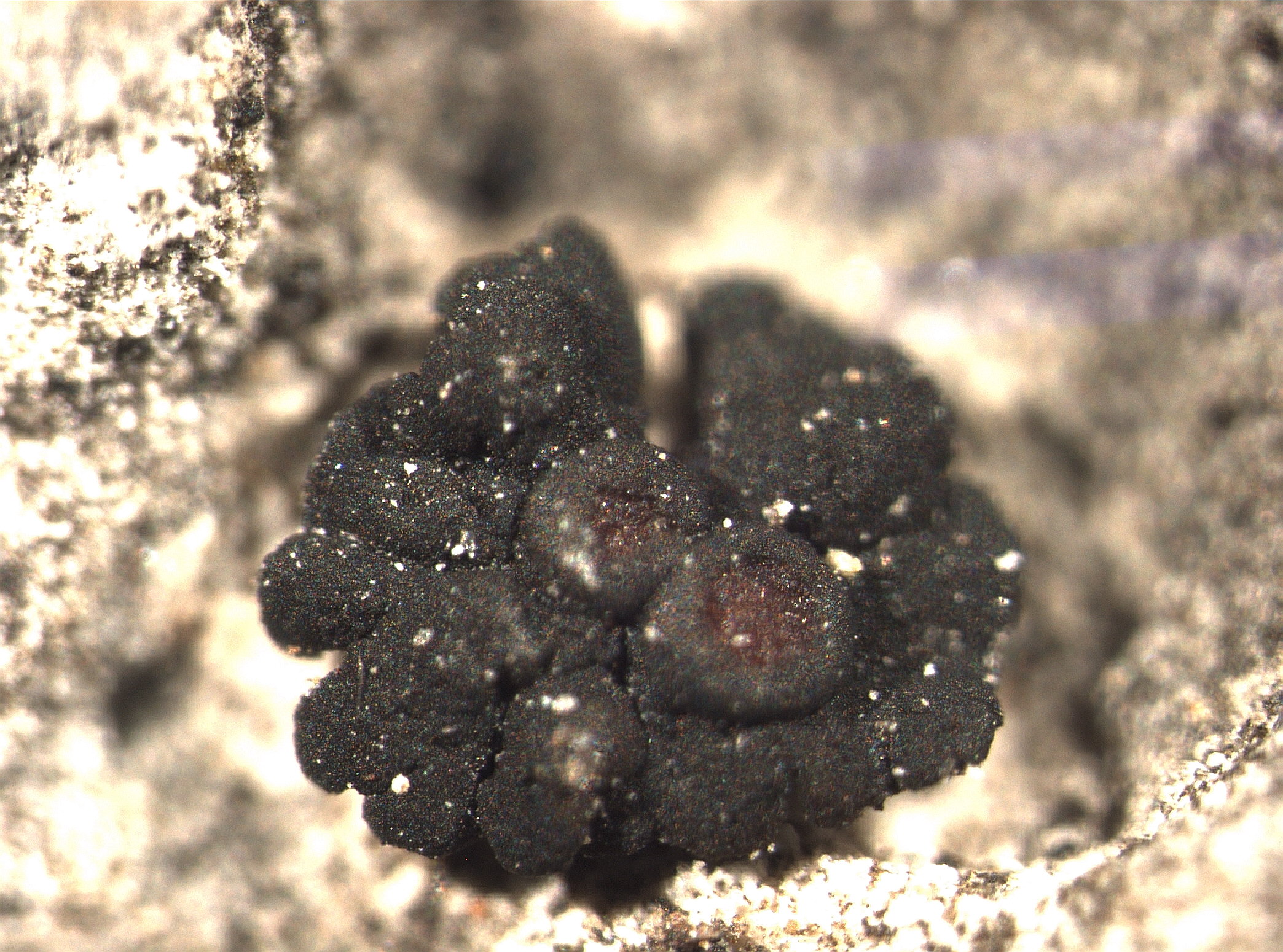
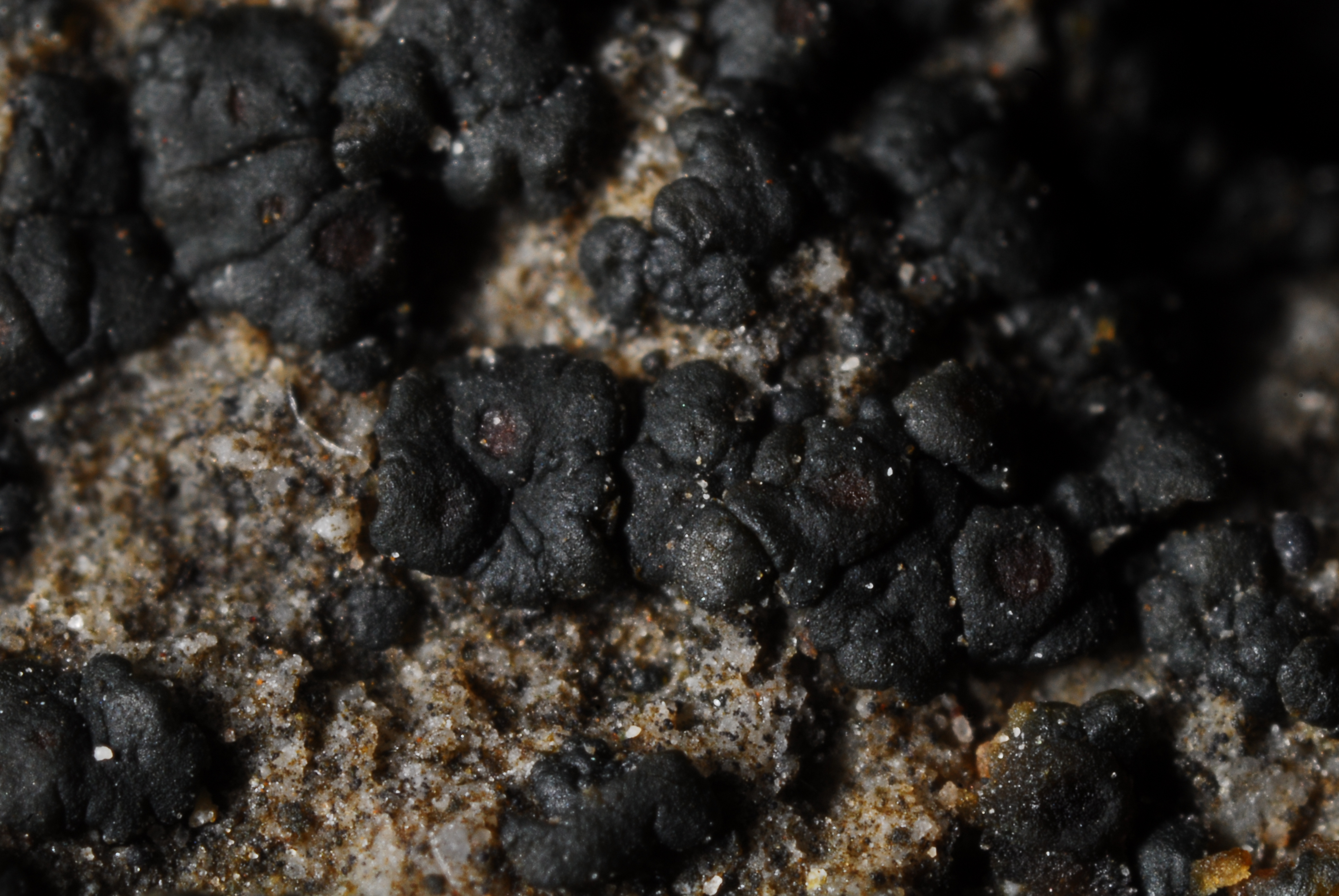
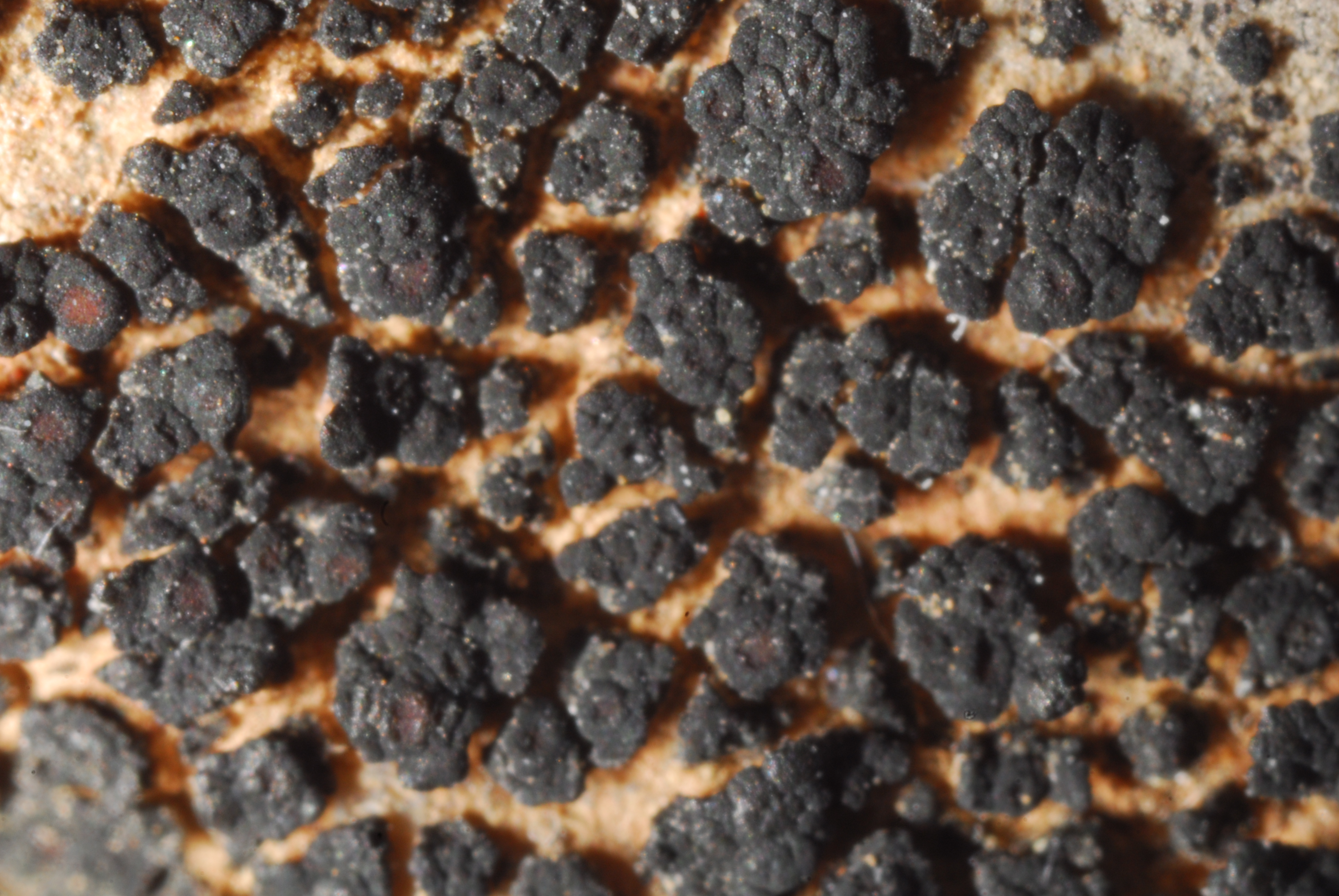
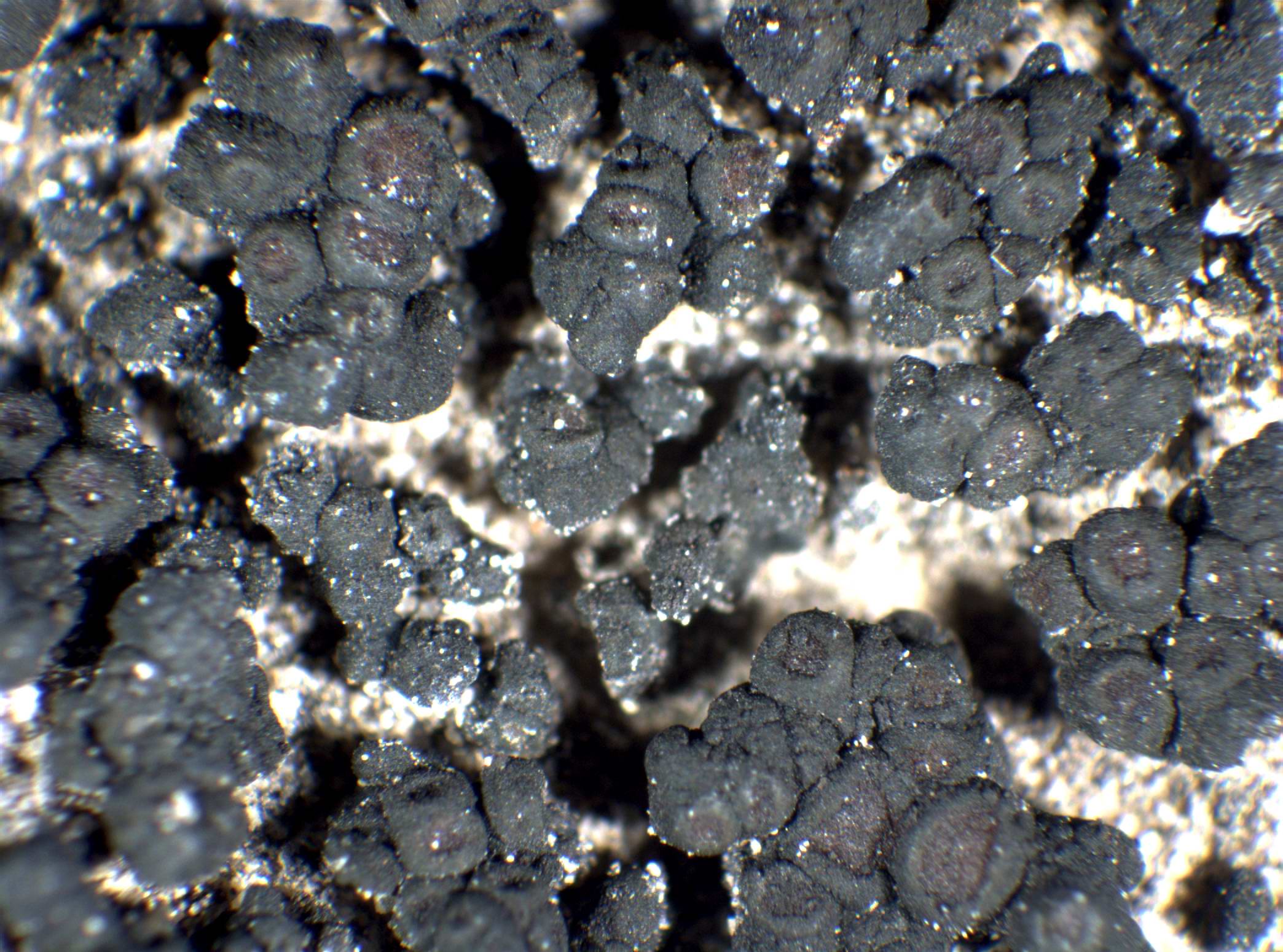
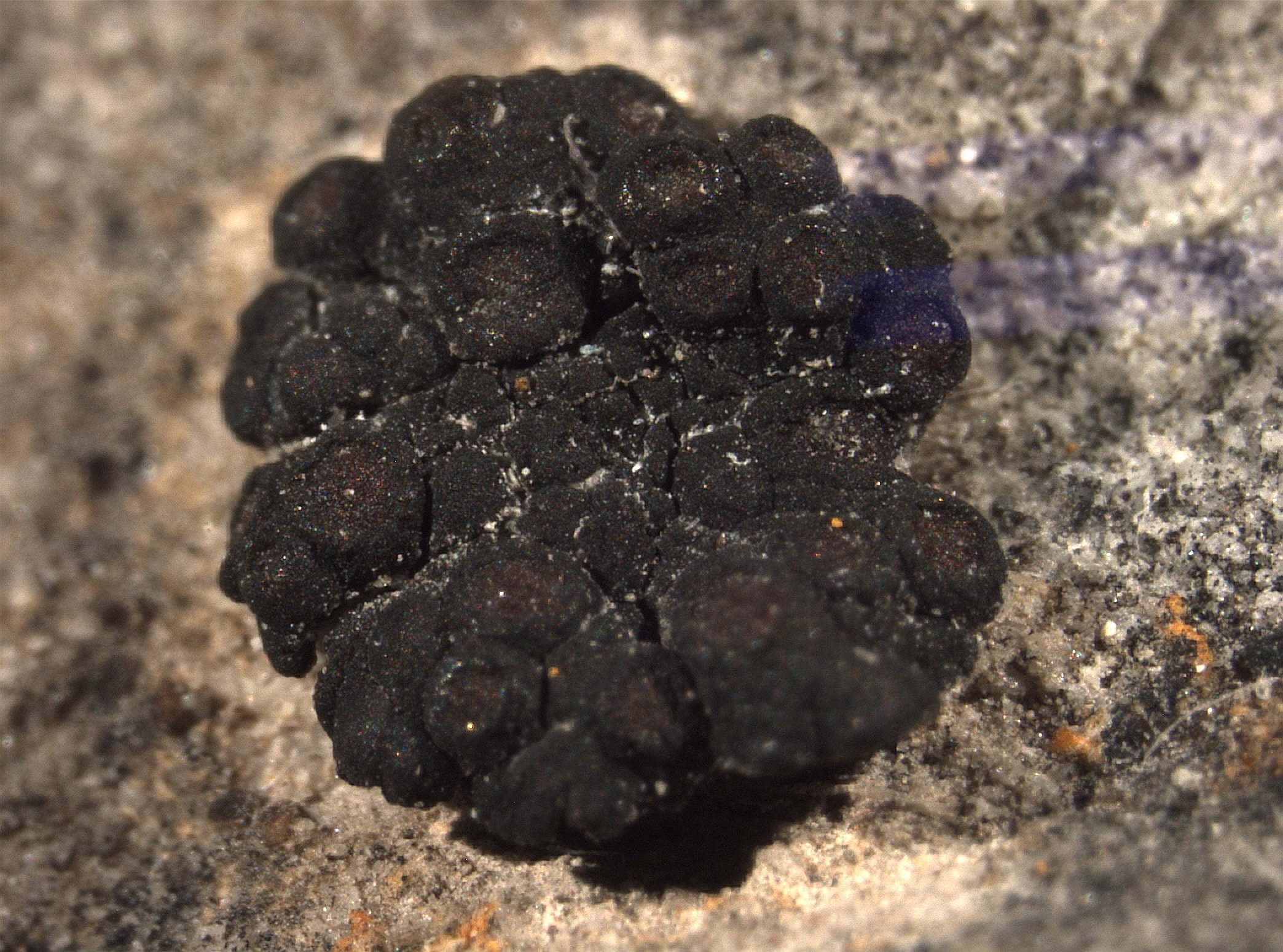
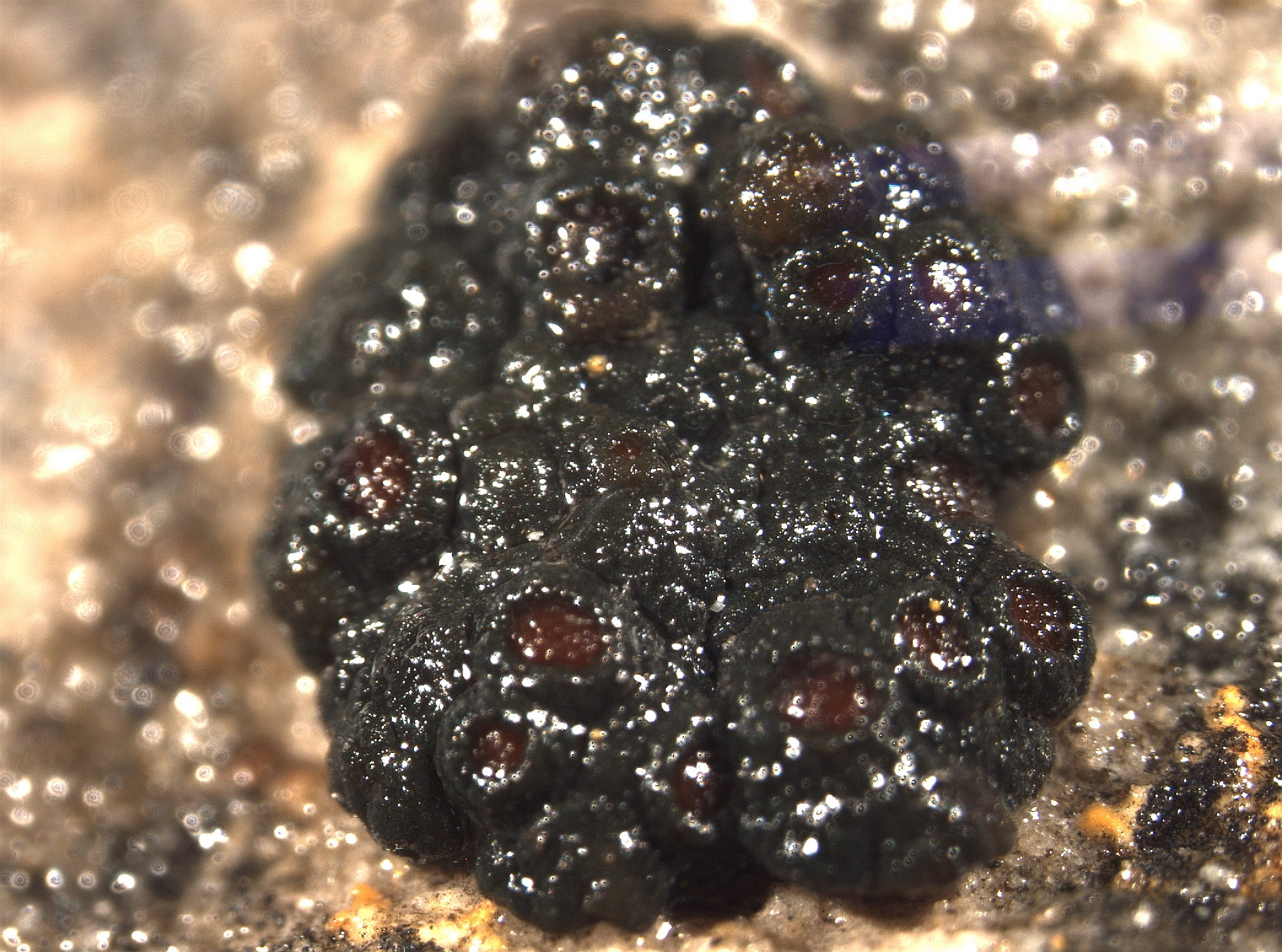
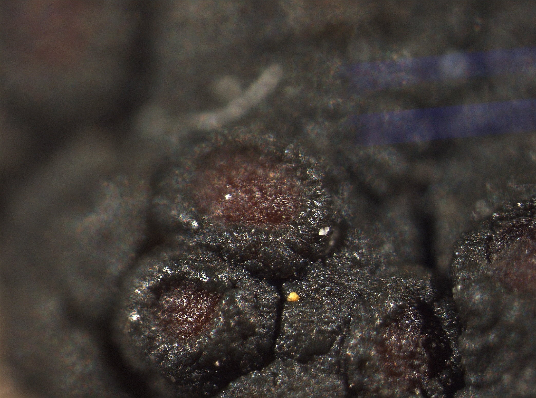
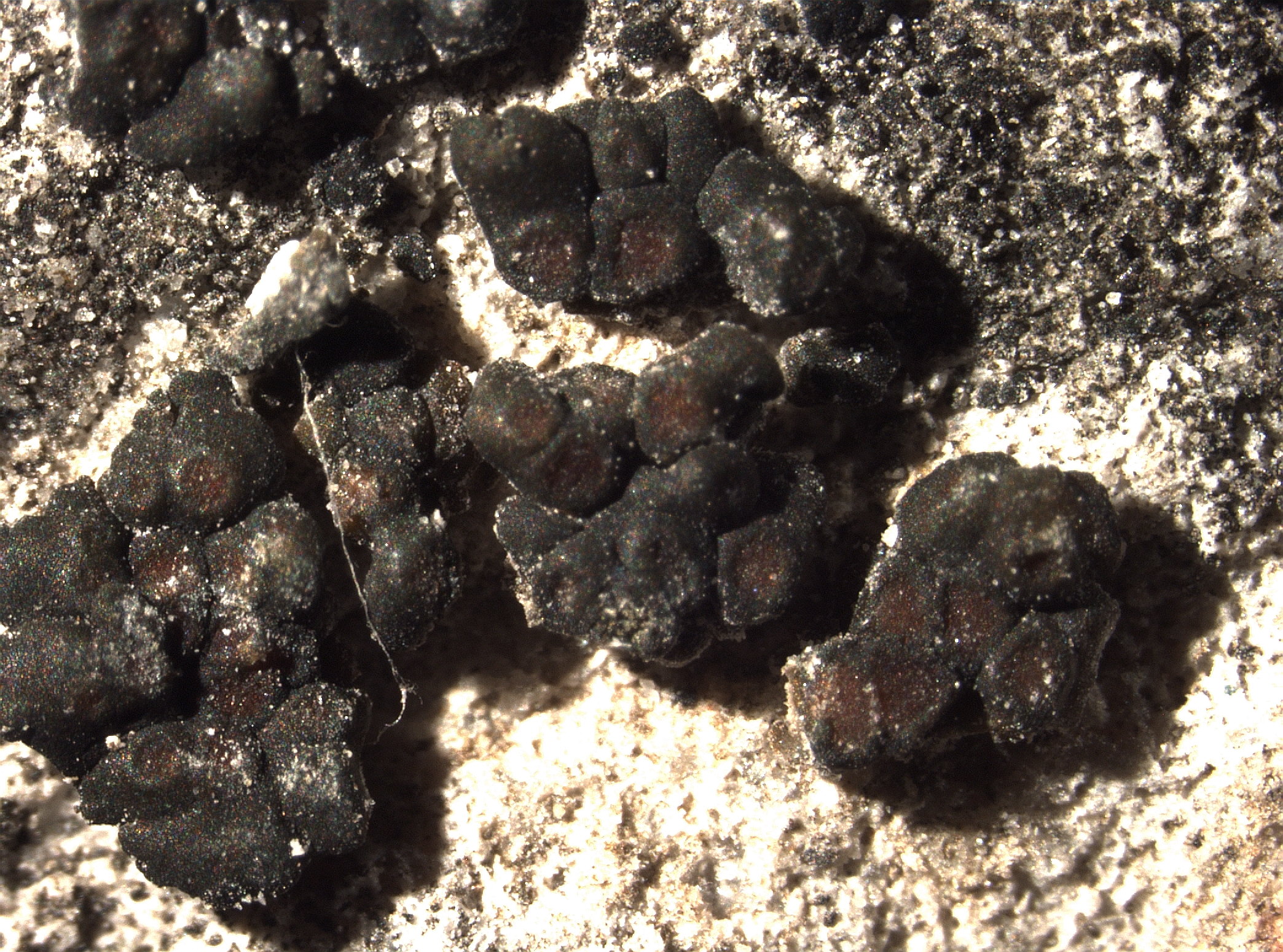
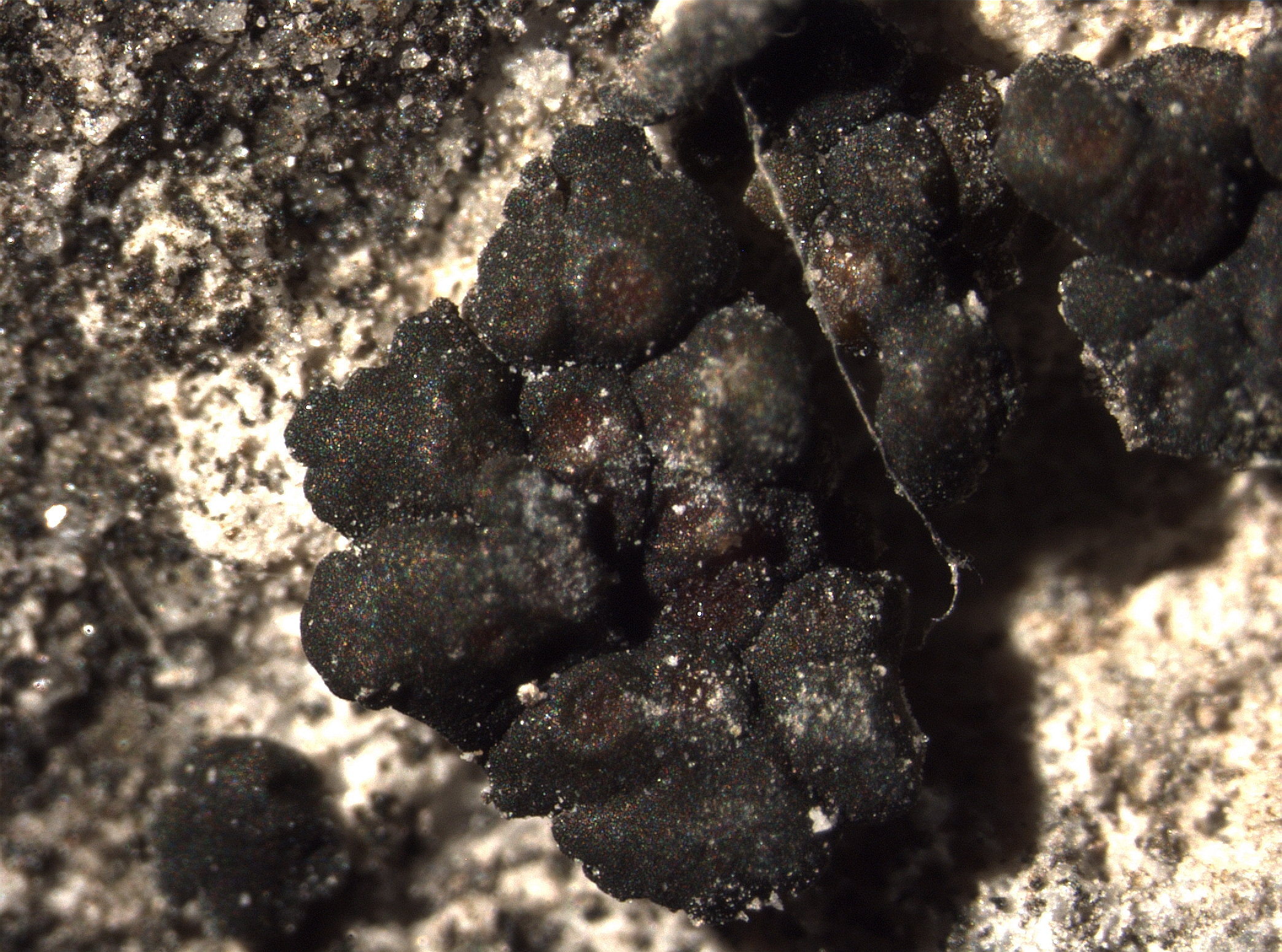
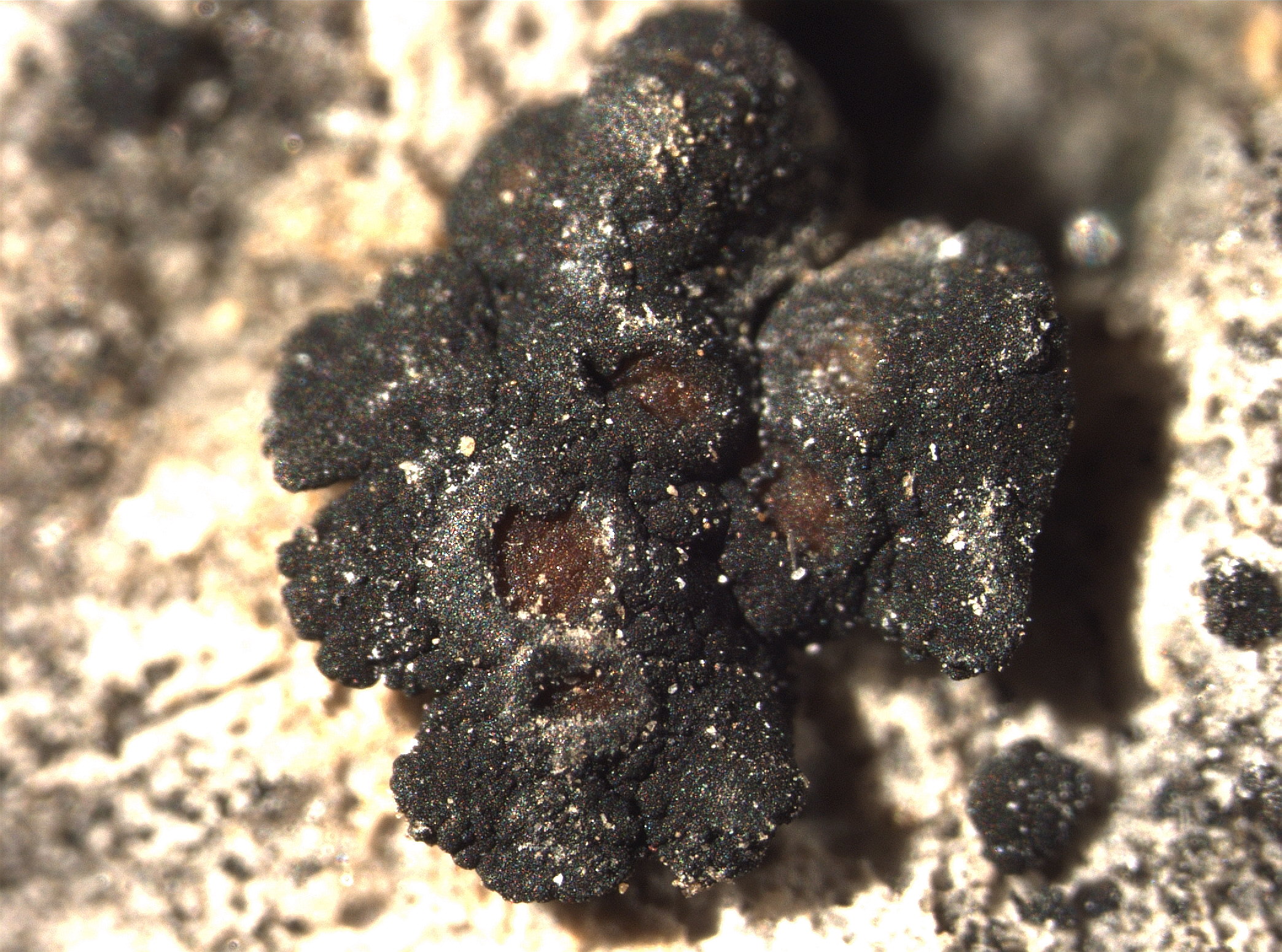
Description: Thallus squamulose-umbilicate, homoiomerous, slightly gelatinous when wet, 0.3-0.45 mm thick, in section with a loosely reticulate network of thin hyphae enclosing groups of cyanobacterial cells. Squamules black, epruinose, dispersed to crowded, closely attached, 5-10(-12) mm wide, button-like, epruinose, not isidiate, attached by a central, compact bundle of rhizohyphae, with a distinctly lobulate margin, the lobules radiating, discrete, diverging and rounded at apex, more or less dichotomously branched, smooth, slightly convex. Apothecia pycnoascocarps, 2-10 or more per squamule, at first immersed and urceolate but soon expanded and clearly lecanorine, 0.3-0.9(-1.2) mm across, with a dark reddish brown, concave to flat disc, and a thick, usually smooth thalline margin. Proper exciple poorly developed; epithecium brownish orange, 5-15 µm high; hymenium colourless, 80-150 µm high, the hymenial gel I+ blue, paraphyses moniliform, 3-4.3 µm thick, the upper cell clearly swollen and to 7 µm wide; hypothecium yellowish, 70-130 µm high. Asci 8-spored, cylindrical-clavate, thin-walled, with an amyloid external apical cap. Ascospores 1-celled, hyaline broadly ellipsoid to ellipsoid, 7-15 x 4-10(-12) µm, the wall 1-2 µm thick. Pycnidia immersed, globose, unilocular. Conidia hyaline, ellipsoid, 2-4 x 0.5-1.5 µm. Photobiont cyanobacterial, chroococcoid, of a few 8-13 µm wide cells often penetrated by haustoria and surrounded by a gelatinous sheath which is colourless in inner parts, yellowish brown near the thallus surface. Spot tests: all negative. Chemistry: without lichen substances. Note: on steeply inclined surfaces of limestone and dolomite with periodical water seepage after rain, below the subalpine belt. Some older records could refer to A. moedligense.
Growth form: Crustose
Substrata: rocks
Photobiont: cyanobacteria, coccaceous (e.g. Gloeocapsa)
Reproductive strategy: mainly sexual
On otherwise dry surfaces with short periods of water seepage after rain
Commonnes-rarity: (info)
Alpine belt: absent
Subalpine belt: absent
Oromediterranean belt: absent
Montane belt: rather rare
Submediterranean belt: common
Padanian area: absent
Humid submediterranean belt: rather common
Humid mediterranean belt: rather rare
Dry mediterranean belt: rather common
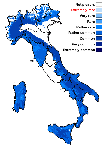
Predictive model
Herbarium samples


Pier Luigi Nimis - Department of Life Sciences, University of Trieste - CC BY-SA 4.0
TSB 2319

Domenico Puntillo; Owner: Domenico Puntillo
Italy

Domenico Puntillo; Owner: Domenico Puntillo
Italy
P.L. Nimis; Owner: Department of Life Sciences, University of Trieste
Herbarium: TSB (9318)
2001/11/21
P.L. Nimis CC BY-SA 4.0
TSB 34297
D. Puntillo CC BY-SA 04

Pier Luigi Nimis - Department of Life Sciences, University of Trieste - CC BY-SA 4.0
TSB 17369
Pier Luigi Nimis - Department of Life Sciences, University of Trieste - CC BY-SA 4.0
TSB 17369
Pier Luigi Nimis - Department of Life Sciences, University of Trieste - CC BY-SA 4.0
TSB 17369

Pier Luigi Nimis - Department of Life Sciences, University of Trieste - CC BY-SA 4.0
TSB 18654

Pier Luigi Nimis - Department of Life Sciences, University of Trieste - CC BY-SA 4.0
TSB 18654
Pier Luigi Nimis - Department of Life Sciences, University of Trieste - CC BY-SA 4.0
TSB 18654
Pier Luigi Nimis - Department of Life Sciences, University of Trieste - CC BY-SA 4.0
TSB 20364
Growth form: Crustose
Substrata: rocks
Photobiont: cyanobacteria, coccaceous (e.g. Gloeocapsa)
Reproductive strategy: mainly sexual
On otherwise dry surfaces with short periods of water seepage after rain
Commonnes-rarity: (info)
Alpine belt: absent
Subalpine belt: absent
Oromediterranean belt: absent
Montane belt: rather rare
Submediterranean belt: common
Padanian area: absent
Humid submediterranean belt: rather common
Humid mediterranean belt: rather rare
Dry mediterranean belt: rather common
Predictive model
| Herbarium samples |
Pier Luigi Nimis - Department of Life Sciences, University of Trieste - CC BY-SA 4.0
TSB 2319
Domenico Puntillo; Owner: Domenico Puntillo
Italy
Domenico Puntillo; Owner: Domenico Puntillo
Italy
P.L. Nimis; Owner: Department of Life Sciences, University of Trieste
Herbarium: TSB (9318)
2001/11/21

P.L. Nimis CC BY-SA 4.0
TSB 34297

D. Puntillo CC BY-SA 04
Pier Luigi Nimis - Department of Life Sciences, University of Trieste - CC BY-SA 4.0
TSB 17369
Pier Luigi Nimis - Department of Life Sciences, University of Trieste - CC BY-SA 4.0
TSB 17369
Pier Luigi Nimis - Department of Life Sciences, University of Trieste - CC BY-SA 4.0
TSB 17369
Pier Luigi Nimis - Department of Life Sciences, University of Trieste - CC BY-SA 4.0
TSB 18654
Pier Luigi Nimis - Department of Life Sciences, University of Trieste - CC BY-SA 4.0
TSB 18654
Pier Luigi Nimis - Department of Life Sciences, University of Trieste - CC BY-SA 4.0
TSB 18654