Sticta fuliginoides Magain & Sérus.
Mycol. Progr., 14, 97: 20, 2015
Synonyms: Sticta fuliginosa auct. Ital. non (Hoffm.) Ach.
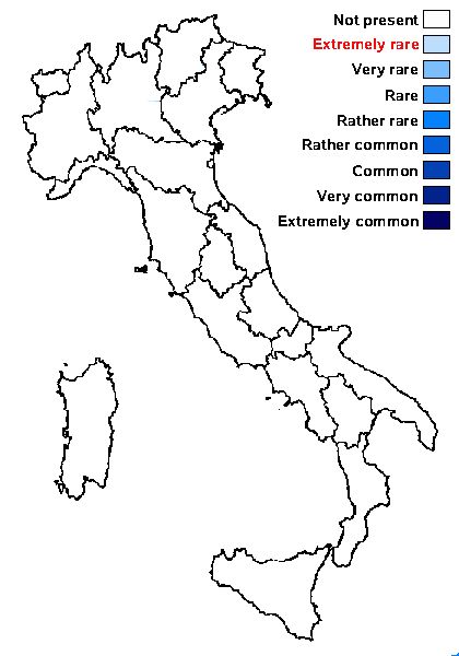
Distribution: N - Frl (Tretiach & Carvalho 1995, Nascimbene & al. 1998, Nascimbene & Caniglia 2003, Tretiach 2004), Ven (Nascimbene 2003b, 2011, Nascimbene & al. 2007, 2010b), TAA (Nascimbene & al. 2007b, 2022, Nimis & al. 2015), Lomb, Piem, VA (Piervittori & Isocrono 1999), Emil (Fariselli & al. 2020). S - Si (Giordani & al. 2009).
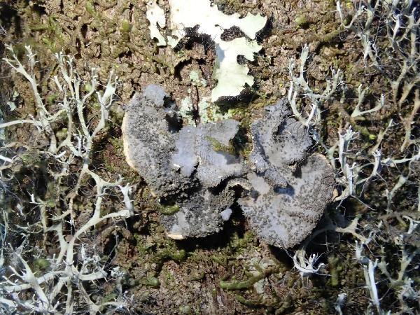
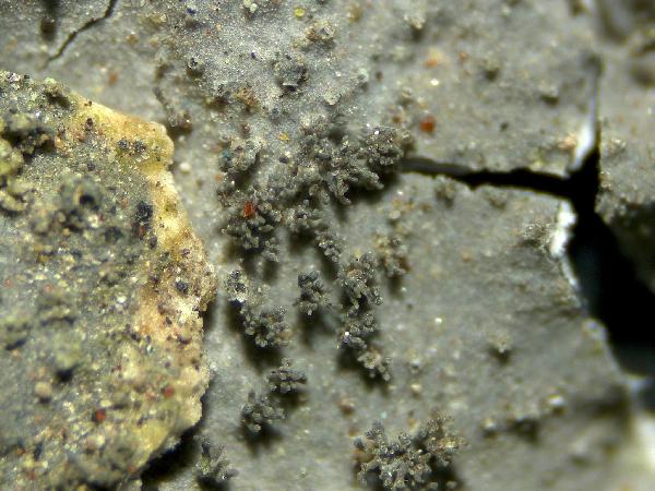
Description: Thallus foliose, heteromerous, dorsiventral, isidiate, at first appearing as a single, vase- or trumpet-shaped lobe with revolute margins, borne on an up to 1 cm long, robust stipe and thus looking like a young mushroom, later developing several flabellate, irregular or mostly suborbicular, 2-3(-5) cm wide lobes. Lobes simple or 1-2 branched, adnate to ascending, especially when young, imbricate and overlapping, flat to slightly involute, with rounded, obtuse or even truncate (especially in old thalli) ends. Upper surface smooth to weakly scrobiculate (especially in old lobes), dark brown or rarely pale grey, dull or slightly glossy. Isidia abundant, laminal, at first simple and globose or with a flattened top, later forming erect coralloid masses, < 0.2 mm high, concolorous with thallus or darker. Lower surface smooth or scrobiculate, pale orange to brownish, darker towards the center in old thalli, sparsely to densely tomentose, with usually abundant, 0.4-0.6(-1.5) mm wide, round or angular, urceolate, erumpent to prominent, white cyphellae whose membrane is made of a layer of rounded cells (5-9 μm in diam.) with outer side (that in contact with the air) covered with minute, 4-6(-8) papillae measuring 1–2.5 x 1-1.5 μm, which tend to disappear in old thalli. Upper cortex paraplectenchymatous, 25-40 μm thick, usually 2-layered, the upper layer made of a single row of smaller cells, the lower layer of 3-5 rows of larger cells; medulla white, lax, 40-60 µm thick; lower cortex paraplectenchymatous, 30-40 μm thick, made of 3–5 rows of isodiametrical cells; primary tomentum agglutinated in fascicles; secondary tomentum always present (high magnification!), made of free, unbranched < 20 μm long, moniliform hyphae, with budding apices producing individual rounded cells that act as diaspores. Apothecia extremely rare (known from a single specimen from Switzerland), lecanorine, laminal, sessile, c. 1 mm across, with a concave, brown disc and a pale orange margin with whitish hairs. Epithecium orange-brown; hymenium brown to red-brown, 60-90 μm high. Asci 8-spored, cylindrical to clavate, unitunicate, with an amyloid cap in the tholus, Peltigera-type. Ascospores 1-3(-7)-septate, hyaline to dark brown, ellipsoid-fusiform, with smooth walls, 16.4-25 x 6.2-8 μm. Photobiont cyanobacterial ( Nostoc, the cells in compact masses). Spot tests: cortex and medulla K-, C-, KC-, P-, UV-. Chemistry: without lichen substances.
Note: a mainly western, Macaronesian-subatlantic species found on mossy trees and rocks within forests or at their edges, incl. parkland conditions in areas with humid, oceanic to suboceanic climates. The species, originally described by Magain & Sérusiaux (2015) and later revised by Sanderson (2016) and Ekman & al. (2019), was never reported from Italy, but, being also known from Switzerland and Central Europe (Bernet & al. 2025), its presence in the Country is probable. Here I follow Bernet & al. (2025) in provisionally attributing most records of the eu-oceanic S. fuliginosa to this species..
Growth form: Foliose, broad lobed
Substrata: bark and rocks
Photobiont: cyanobacteria, filamentous (e.g. Nostoc, Scytonema)
Reproductive strategy: mainly asexual, by isidia, or isidia-like structures (e.g. schizidia)
Restricted to humid-warm, oceanic areas
Commonnes-rarity: (info)
Alpine belt: absent
Subalpine belt: absent
Oromediterranean belt: absent
Montane belt: very rare
Submediterranean belt: absent
Padanian area: absent
Humid submediterranean belt: extremely rare
Humid mediterranean belt: absent
Dry mediterranean belt: absent
Predictive model
Herbarium samples

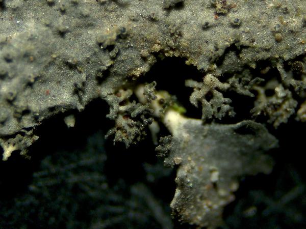
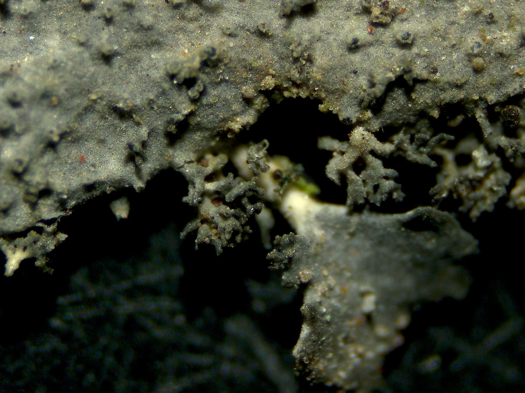
Image uploaded by Paul Cannon - CC BY-SA NC - Source: https://fungi.myspecies.info/all-fungi/sticta-fuliginoides
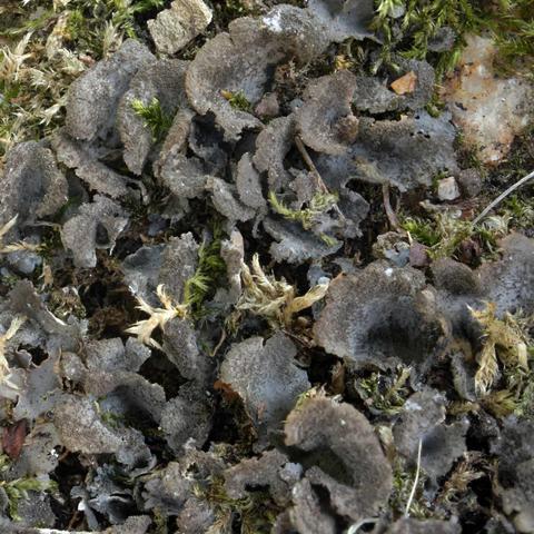
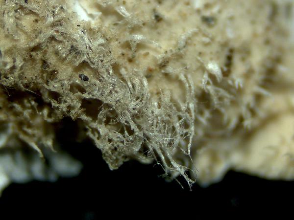
Image uploaded by Paul Cannon - CC BY-SA NC - Source: https://fungi.myspecies.info/all-fungi/sticta-fuliginoides
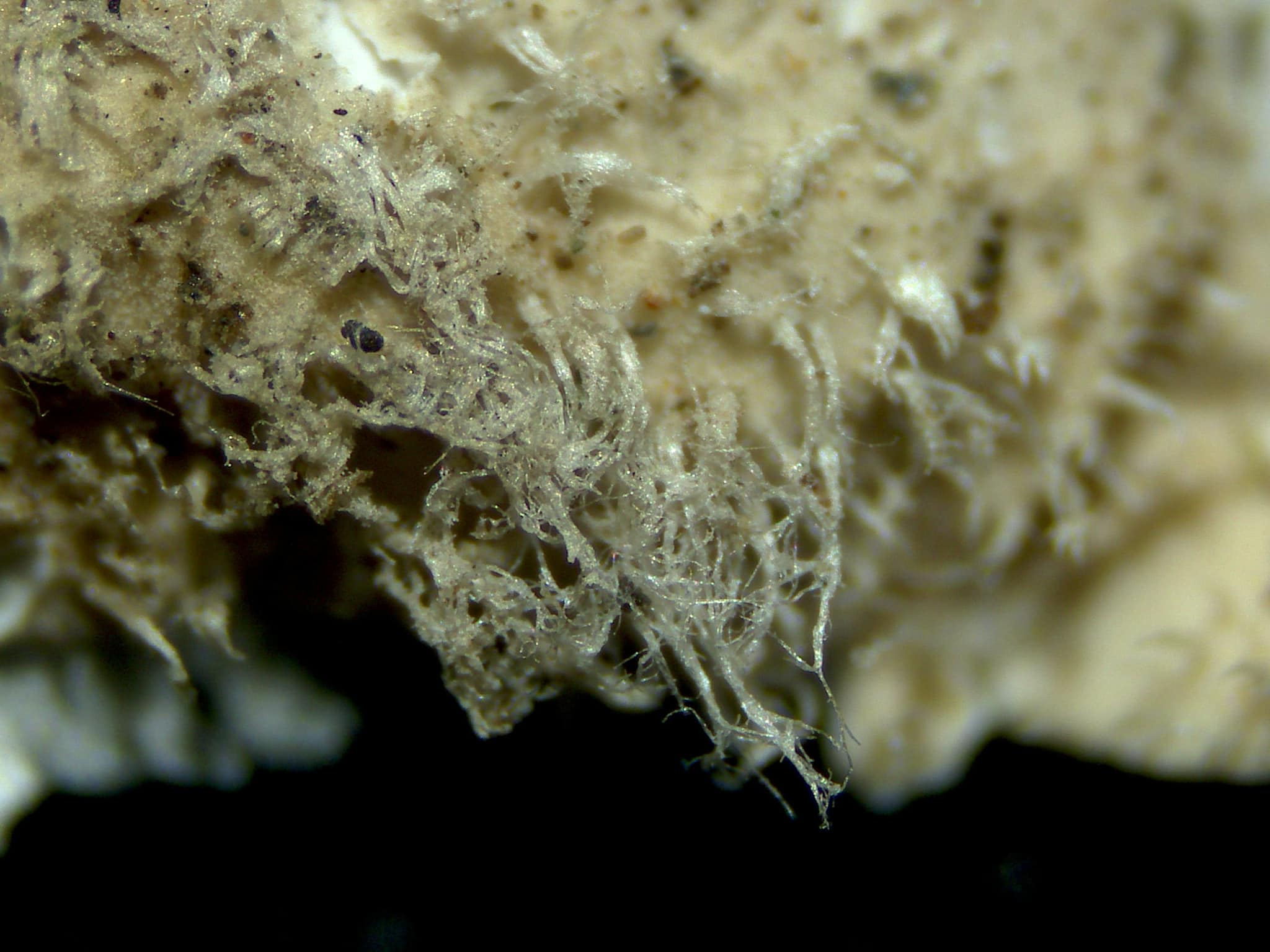
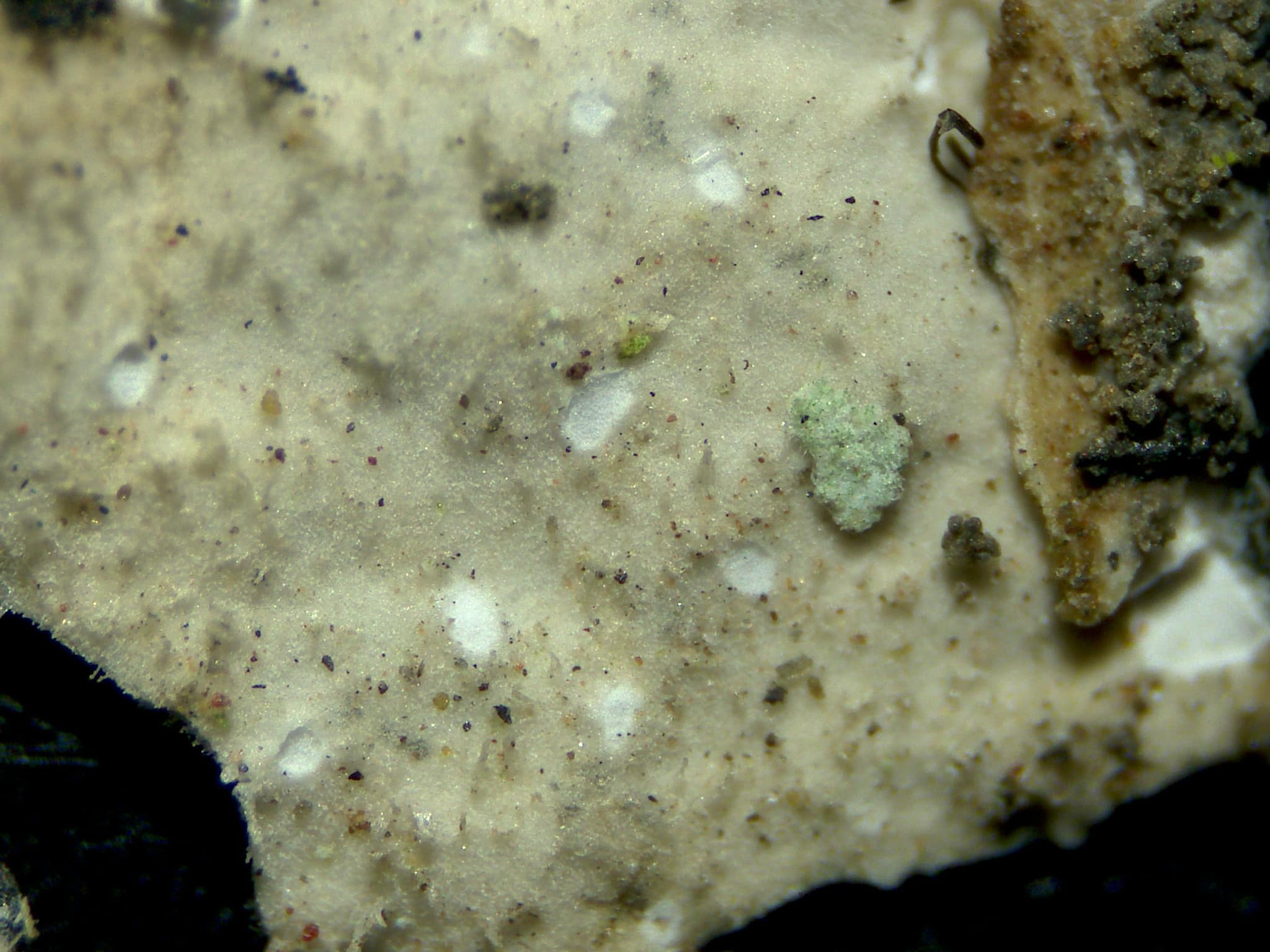
Manuel Gil
Tenerife. Canary Islands Corticolous
Manuel Gil
Tenerife. Canary Islands Corticolous
Manuel Gil
Tenerife. Canary Islands Corticolous
Manuel Gil
Tenerife. Canary Islands Corticolous
Manuel Gil
Tenerife. Canary Islands Corticolous
Manuel Gil
Tenerife. Canary Islands Corticolous
Manuel Gil
Tenerife. Canary Islands Corticolous
Manuel Gil
Tenerife. Canary Islands Corticolous
Manuel Gil
Tenerife. Canary Islands Corticolous
Manuel Gil
Tenerife. Canary Islands Corticolous
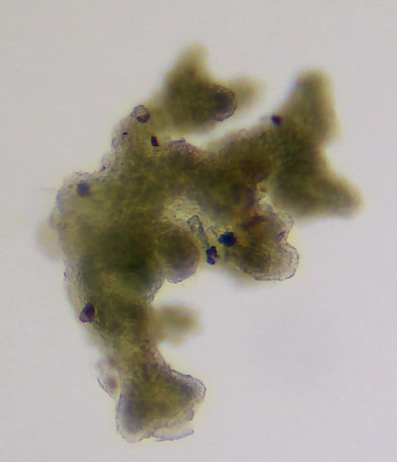
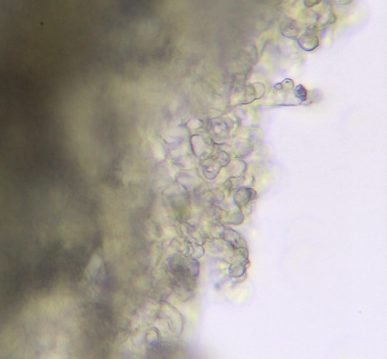
Source: Ekman, S., Tønsberg, T. & Jørgensen, P. M. 2019. The Sticta fuliginosa group in Norway and Sweden.Graphis Scripta 31 (4): 23–33. Oslo. ISSN 2002-4495. - CC BY-4.0
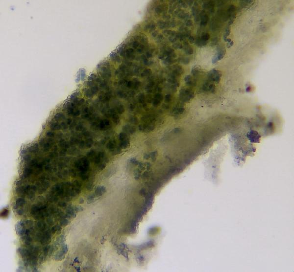
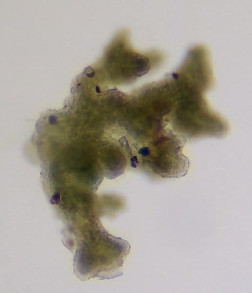
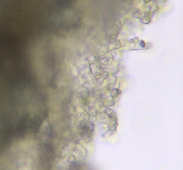
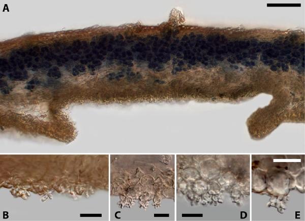
Sections through cyphellae in Sticta. A–E. S. fuliginoides, showing section through entire cyphella(A) and cyphella surface cells with papillae (B–E). Scales: 0.1 mm (A), 10 μm (B–E). Photos of UPS L-112257 (A), L- 157135 (B), L-002176 (C–E)
Source: Ekman, S., Tønsberg, T. & Jørgensen, P. M. 2019. The Sticta fuliginosa group in Norway and Sweden.Graphis Scripta 31 (4): 23–33. Oslo. ISSN 2002-4495. - CC BY-4.0
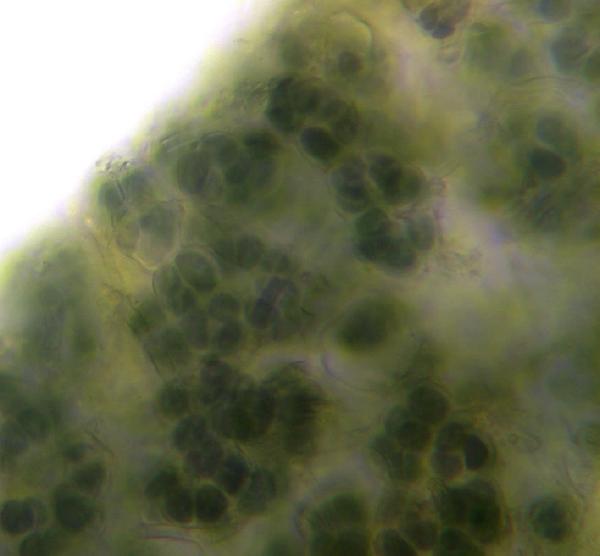
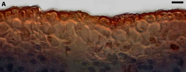
Sections through upper cortex in Sticta. A. S. fuliginoides. Scales: 10 μm. Photo of UPS L-133433
Growth form: Foliose, broad lobed
Substrata: bark and rocks
Photobiont: cyanobacteria, filamentous (e.g. Nostoc, Scytonema)
Reproductive strategy: mainly asexual, by isidia, or isidia-like structures (e.g. schizidia)
Restricted to humid-warm, oceanic areas
Commonnes-rarity: (info)
Alpine belt: absent
Subalpine belt: absent
Oromediterranean belt: absent
Montane belt: very rare
Submediterranean belt: absent
Padanian area: absent
Humid submediterranean belt: extremely rare
Humid mediterranean belt: absent
Dry mediterranean belt: absent
Predictive model
| Herbarium samples |
 DOLICHENS
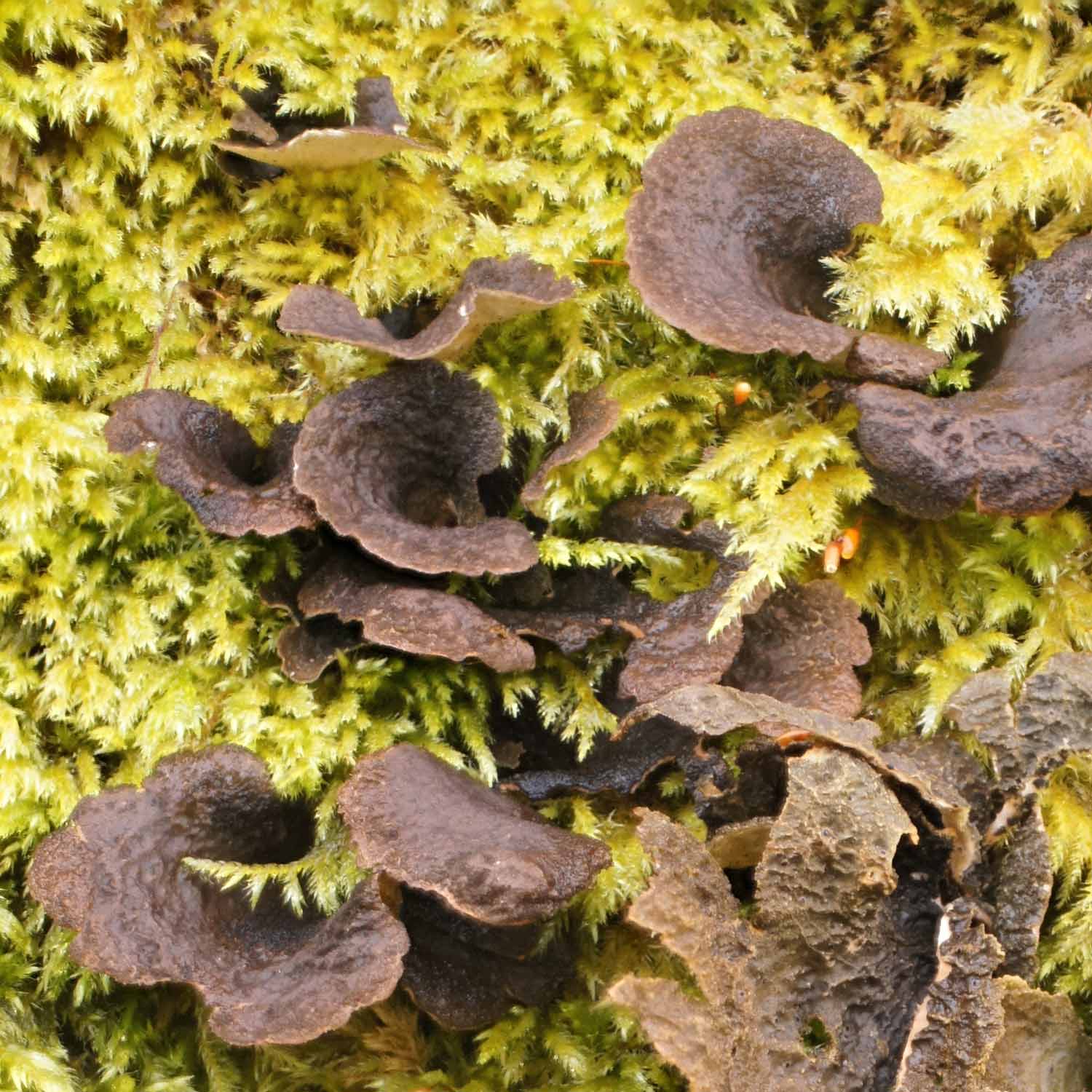
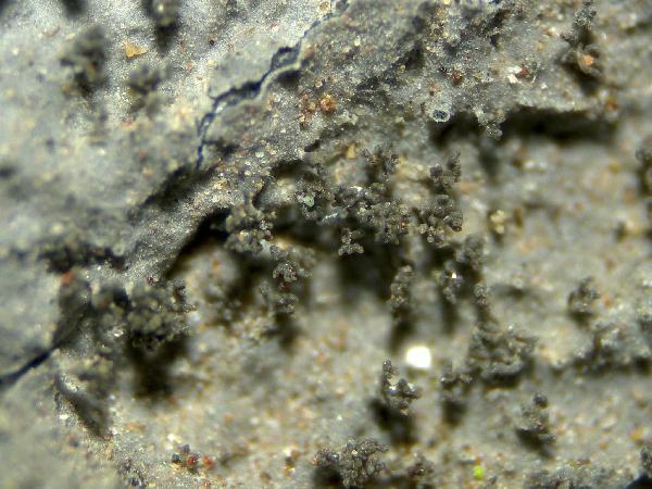
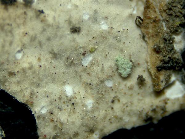
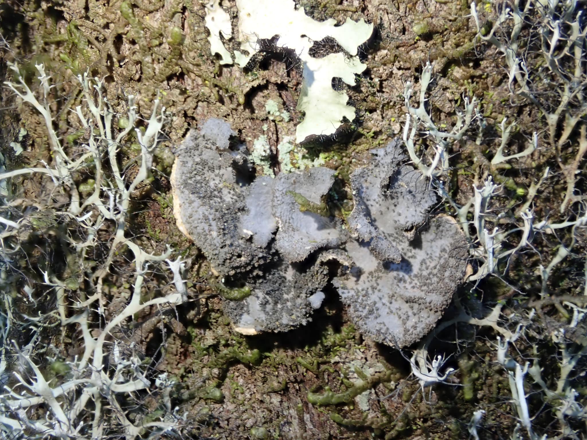
Image uploaded by Paul Cannon - CC BY-SA NC - Source: https://fungi.myspecies.info/all-fungi/sticta-fuliginoides

Image uploaded by Paul Cannon - CC BY-SA NC - Source: https://fungi.myspecies.info/all-fungi/sticta-fuliginoides
Manuel Gil
Tenerife. Canary Islands Corticolous

Manuel Gil
Tenerife. Canary Islands Corticolous
Manuel Gil
Tenerife. Canary Islands Corticolous
Manuel Gil
Tenerife. Canary Islands Corticolous

Manuel Gil
Tenerife. Canary Islands Corticolous
Manuel Gil
Tenerife. Canary Islands Corticolous

Manuel Gil
Tenerife. Canary Islands Corticolous
Manuel Gil
Tenerife. Canary Islands Corticolous
Manuel Gil
Tenerife. Canary Islands Corticolous

Manuel Gil
Tenerife. Canary Islands Corticolous

Source: Ekman, S., Tønsberg, T. & Jørgensen, P. M. 2019. The Sticta fuliginosa group in Norway and Sweden.Graphis Scripta 31 (4): 23–33. Oslo. ISSN 2002-4495. - CC BY-4.0
Sections through cyphellae in Sticta. A–E. S. fuliginoides, showing section through entire cyphella(A) and cyphella surface cells with papillae (B–E). Scales: 0.1 mm (A), 10 μm (B–E). Photos of UPS L-112257 (A), L- 157135 (B), L-002176 (C–E)