Circinaria viridescens (A. Massal.)
Provisionally placed here, ICN Art. 36.1b.. Basionym: Pachyospora viridescens A. Massal. - Ric. Auton. Lich. Crost.: 46, 1852.
Synonyms: Aspicilia viridescens (A. Massal.) Hue
Distribution: N - Ven (Massalongo 1852), TAA (Dalla Torre & Sarnthein 1902), Emil (Fariselli & al. 2020), Lig (Giordani & al. 2016). C - Laz (Nimis & Tretiach 2004), Abr (Nimis & Tretiach 1999, Gheza & al. 2021), Mol (Nimis & Tretiach 1999, Caporale & al. 2008), Sar (Rizzi & al. 2011, Giordani & al. 2013). S - Camp (Nimis & Tretiach 2004), Pugl (Nimis & Tretiach 1999), Bas (Nimis & Tretiach 1999), Cal (Puntillo 2011), Si (Nimis & al. 1994, 1996b, Ottonello & al. 1994, Ottonello & Romano 1997, Grillo & al. 2007).
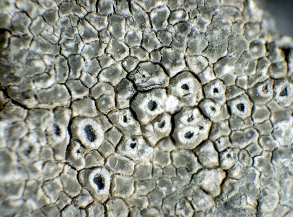
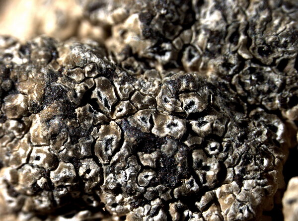
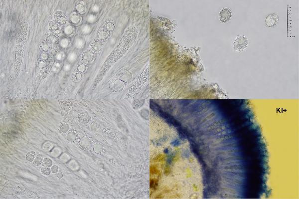
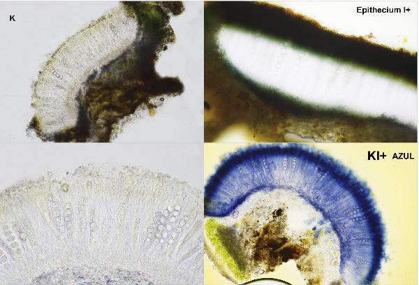
Description: Thallus crustose, episubstratic, areolate, glaucous green, forming regular, usually well-delimited patches, the areoles angular or irregular, concave with slightly raised margins and sometimes subumbonate in the center (primordium of apothecium), 0.3-1 mm wide, contiguous and separated by cracks. Cortex paraplectenchymatous, the upper part brownish green, the pigment N+ emerald green, K+ yellow-brown (Caesiocinerea-green pigment). Apothecia aspicilioid, 0.2-0.5(-0.8) mm across, immersed in the areoles, 1(-3) per areole, at first crateriform, later with an expanded disc, round or angular, with a black, sometimes weakly pruinose, concave to flat disc and a distinctly raised, often white-pruinose thalline margin. Proper exciple poorly developed, colourless; epithecium olive green to olive-brown, with crystals, N+ green, K+ yellow-brown; hymenium colourless, I+ blue partly turning yellow-brown; paraphyses slightly branched and anastomosing, 1.5-2.5 µm thick at mid-level, submoniliform in upper part, the uppermost cells subglobose, (3-)4-6 µm wide; subhymenium and hypothecium colourless. Asci (2-)4(-6)-spored, clavate, the thin outer coat K/I+ blue, the wall and apical dome K/I-. Ascospores 1-celled, hyaline, globose to subglobose, 16-35 x 14-29 µm. Pycnidia rare, immersed, with a black, punctiform ostiole and a colourless wall. Conidia thread-like, straight or slightly curved, 8-12 x c. 1 µm. Photobiont chlorococcoid. Spot tests: cortex and medulla K-, C-, KC-, P-. Chemistry: usually with aspicilin.Note: on base-rich, hard siliceous rocks (e.g. basalt), most frequent in Southern Italy. Several south Italian records of Aspicilia contorta s.lat. reported by Nimis (1993: 100) could refer to this taxon, which, however, can be easily confused with grey-green morphotypes of C. hoffmanniana that grows on more or less calciferous substrata (see Roux & coll. 2014, 2020).
Growth form: Crustose
Substrata: rocks
Photobiont: green algae other than Trentepohlia
Reproductive strategy: mainly sexual
Most common in areas with a humid-warm climate (e.g. most of Tyrrenian Italy)
Commonnes-rarity: (info)
Alpine belt: absent
Subalpine belt: absent
Oromediterranean belt: absent
Montane belt: very rare
Submediterranean belt: rare
Padanian area: absent
Humid submediterranean belt: rather common
Humid mediterranean belt: rather rare
Dry mediterranean belt: extremely rare
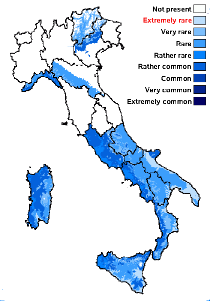
Predictive model
Herbarium samples
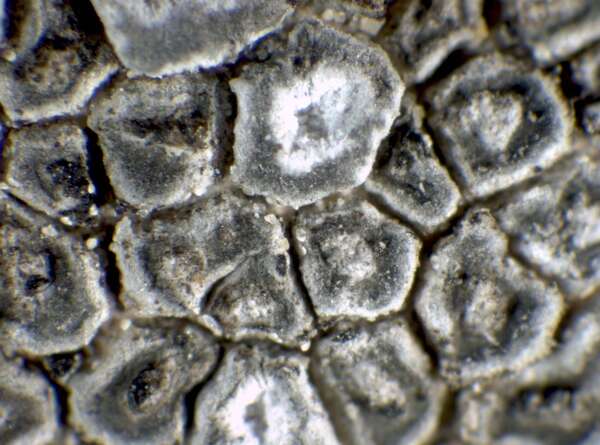

P.L. Nimis; Owner: Department of Life Sciences, University of Trieste
Herbarium: TSB (22740)
2001/12/09
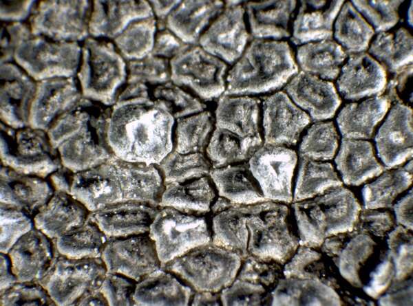

P.L. Nimis; Owner: Department of Life Sciences, University of Trieste
Herbarium: TSB (22740)
2001/12/09
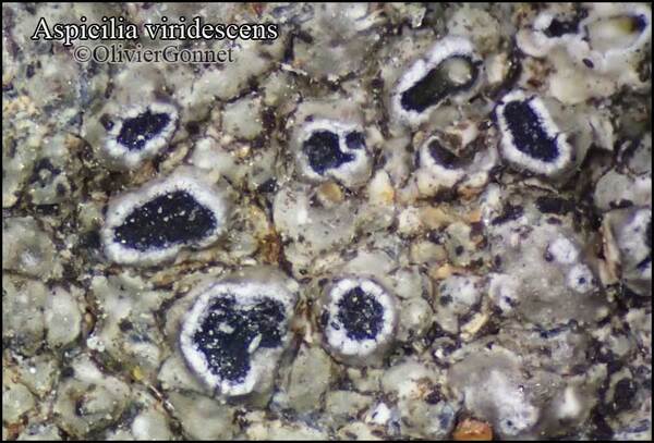
Courtesy Danièle et Olivier Gonnet - Source: https://www.afl-lichenologie.fr/Photos_AFL/Photos_AFL_A/Aspicilia_viridescens.htm
France, Stage micro de nov. 2015 - Saint-Prejet-Armandon - Haute-Loire
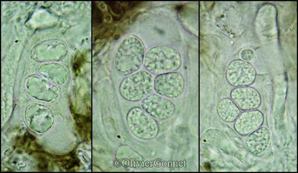
Courtesy Danièle et Olivier Gonnet - Source: https://www.afl-lichenologie.fr/Photos_AFL/Photos_AFL_A/Aspicilia_viridescens.htm
France, Stage micro de nov. 2015 - Saint-Prejet-Armandon - Haute-Loire
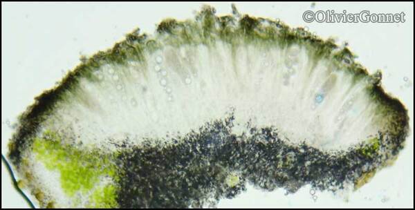
Courtesy Danièle et Olivier Gonnet - Source: https://www.afl-lichenologie.fr/Photos_AFL/Photos_AFL_A/Aspicilia_viridescens.htm
France, Stage micro de nov. 2015 - Saint-Prejet-Armandon - Haute-Loire
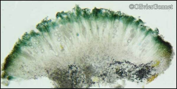
Courtesy Danièle et Olivier Gonnet - Source: https://www.afl-lichenologie.fr/Photos_AFL/Photos_AFL_A/Aspicilia_viridescens.htm
France, Stage micro de nov. 2015 - Saint-Prejet-Armandon - Haute-Loire
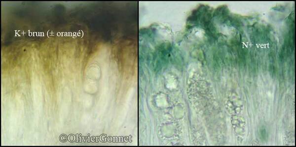
Courtesy Danièle et Olivier Gonnet - Source: https://www.afl-lichenologie.fr/Photos_AFL/Photos_AFL_A/Aspicilia_viridescens.htm
France, Stage micro de nov. 2015 - Saint-Prejet-Armandon - Haute-Loire
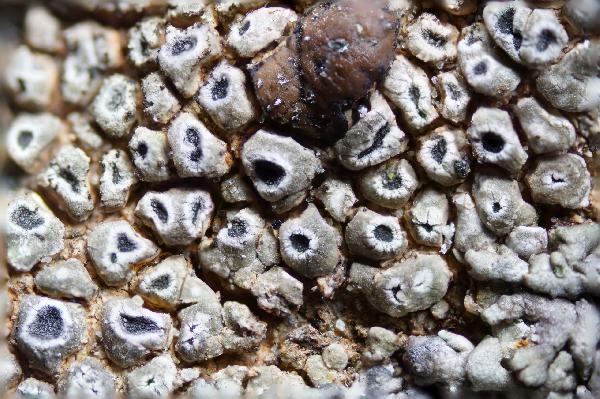
Marta Gonzalez Garcia - Centro de Estudios Micologicos Asturianos
Spain, Braña del Puerto de la Mesa (Somiedo-Asturias), 24-X-2021, sobre rocas silíceas junto a Protoparmeliopsis bolcana, y Acarospora veronensis, leg. & det. M. González, ERD-9176.
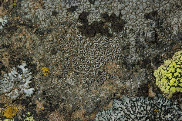
Marta Gonzalez Garcia - Centro de Estudios Micologicos Asturianos
Spain, Braña del Puerto de la Mesa (Somiedo-Asturias), 24-X-2021, sobre rocas silíceas junto a Protoparmeliopsis bolcana, y Acarospora veronensis, leg. & det. M. González, ERD-9176.
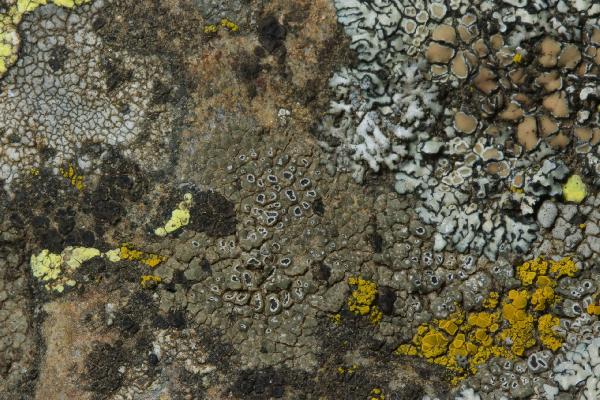
Marta Gonzalez Garcia - Centro de Estudios Micologicos Asturianos
Spain, Braña del Puerto de la Mesa (Somiedo-Asturias), 24-X-2021, sobre rocas silíceas junto a Protoparmeliopsis bolcana, y Acarospora veronensis, leg. & det. M. González, ERD-9176.
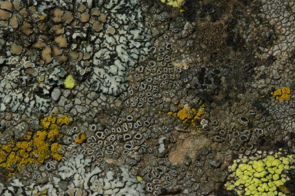
Marta Gonzalez Garcia - Centro de Estudios Micologicos Asturianos
Spain, Braña del Puerto de la Mesa (Somiedo-Asturias), 24-X-2021, sobre rocas silíceas junto a Protoparmeliopsis bolcana, y Acarospora veronensis, leg. & det. M. González, ERD-9176.
Growth form: Crustose
Substrata: rocks
Photobiont: green algae other than Trentepohlia
Reproductive strategy: mainly sexual
Most common in areas with a humid-warm climate (e.g. most of Tyrrenian Italy)
Commonnes-rarity: (info)
Alpine belt: absent
Subalpine belt: absent
Oromediterranean belt: absent
Montane belt: very rare
Submediterranean belt: rare
Padanian area: absent
Humid submediterranean belt: rather common
Humid mediterranean belt: rather rare
Dry mediterranean belt: extremely rare

Predictive model
| Herbarium samples |


P.L. Nimis; Owner: Department of Life Sciences, University of Trieste
Herbarium: TSB (22740)
2001/12/09


P.L. Nimis; Owner: Department of Life Sciences, University of Trieste
Herbarium: TSB (22740)
2001/12/09

Courtesy Danièle et Olivier Gonnet - Source: https://www.afl-lichenologie.fr/Photos_AFL/Photos_AFL_A/Aspicilia_viridescens.htm
France, Stage micro de nov. 2015 - Saint-Prejet-Armandon - Haute-Loire

Courtesy Danièle et Olivier Gonnet - Source: https://www.afl-lichenologie.fr/Photos_AFL/Photos_AFL_A/Aspicilia_viridescens.htm
France, Stage micro de nov. 2015 - Saint-Prejet-Armandon - Haute-Loire

Courtesy Danièle et Olivier Gonnet - Source: https://www.afl-lichenologie.fr/Photos_AFL/Photos_AFL_A/Aspicilia_viridescens.htm
France, Stage micro de nov. 2015 - Saint-Prejet-Armandon - Haute-Loire

Courtesy Danièle et Olivier Gonnet - Source: https://www.afl-lichenologie.fr/Photos_AFL/Photos_AFL_A/Aspicilia_viridescens.htm
France, Stage micro de nov. 2015 - Saint-Prejet-Armandon - Haute-Loire

Courtesy Danièle et Olivier Gonnet - Source: https://www.afl-lichenologie.fr/Photos_AFL/Photos_AFL_A/Aspicilia_viridescens.htm
France, Stage micro de nov. 2015 - Saint-Prejet-Armandon - Haute-Loire

Marta Gonzalez Garcia - Centro de Estudios Micologicos Asturianos
Spain, Braña del Puerto de la Mesa (Somiedo-Asturias), 24-X-2021, sobre rocas silíceas junto a Protoparmeliopsis bolcana, y Acarospora veronensis, leg. & det. M. González, ERD-9176.

Marta Gonzalez Garcia - Centro de Estudios Micologicos Asturianos
Spain, Braña del Puerto de la Mesa (Somiedo-Asturias), 24-X-2021, sobre rocas silíceas junto a Protoparmeliopsis bolcana, y Acarospora veronensis, leg. & det. M. González, ERD-9176.

Marta Gonzalez Garcia - Centro de Estudios Micologicos Asturianos
Spain, Braña del Puerto de la Mesa (Somiedo-Asturias), 24-X-2021, sobre rocas silíceas junto a Protoparmeliopsis bolcana, y Acarospora veronensis, leg. & det. M. González, ERD-9176.

 DOLICHENS
